
01
Infections
Bacteria, viruses, fungi or acanthamoeba can invade the cornea, especially after injury or contact-lens wear, and produce ulceration.
Clear Vision. Expert Care. Healthier Eyes.
M G Eye Foundation/Services/Cornea Services
M G Eye Foundation, Bara Gamharia, Jamshedpur, provides assessment and treatment for corneal abrasion, ulcer, scarring, keratoconus and other diseases of the clear front window of the eye. DR KUMAR SAKET and DR MALAY KUMAR DWIVEDI plan care after a detailed examination of the ocular surface.
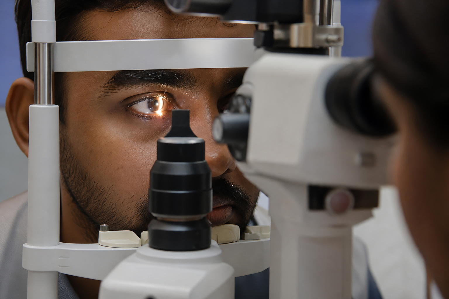
The cornea is the clear, curved front surface of the eye. It focuses light and protects deeper structures. Infection, injury, dystrophy, thinning or dry eye can cloud or distort this surface. When the cornea is damaged, vision may fall even if the lens and retina are healthy.
Why it develops
Corneal disease has many starting points. Infection and injury are common; dystrophy and thinning may run in families.
Bacteria, viruses, fungi or acanthamoeba can invade the cornea, especially after injury or contact-lens wear, and produce ulceration.
A scratch, foreign body or blunt injury can break the epithelial barrier and allow infection or scarring.
Chemical splash, thermal burn or penetrating injury may destroy corneal tissue and need urgent washing and specialist care.
Alkali and acid exposure damage cells rapidly. Immediate irrigation and examination reduce the depth of injury.
Inherited or age-related change in corneal layers can cause haze, recurrent erosion or progressive thinning.
Systemic immune disease may inflame the ocular surface or sclera and secondarily involve the cornea.
Keratoconus and related ectasias steepen and thin the cornea, distorting vision and sometimes progressing.
Severe vitamin A deficiency and related malnutrition can soften the cornea and raise the risk of ulcer.
Chronic allergic inflammation roughens the surface, causes itching and may lead to shield ulcers in children.
Benign or malignant growths of the ocular surface can invade or distort the cornea.
A poor tear film or long-term pressure disease and its drops can stress the surface and delay healing.
How it feels
Surface disease often hurts. Seek care quickly if pain, discharge or sudden blur follows injury or lens wear.
01
The cornea has many nerves. Even a small abrasion can cause sharp pain, especially on blinking.
02
Mucoid or purulent discharge suggests infection and needs examination the same day when possible.
03
Haze, irregular astigmatism or oedema scatter light so print and faces lose sharpness.
04
Reflex watering is the eye’s attempt to wash an irritated surface.
05
Ciliary injection around the cornea is a warning sign of keratitis or ulcer.
06
Photophobia is typical when the epithelium is broken or inflamed.
07
After healing, a white opacity may remain and permanently reduce vision if it sits on the visual axis.
Mild injuries may be treated with a patch, a bandage contact lens, prescribed drops or tablets. Advanced disease such as dense scar, failed endothelium or uncontrolled infection may need corneal transplant or other surgery. Keratoconus is often managed first with rigid or scleral lenses. Autoimmune disease is treated together with the underlying systemic condition. Cross-linking (CXL) is used to slow progressive ectasia when the cornea is suitable.
01
A full-thickness transplant removes the diseased central cornea and replaces it with donor tissue. It is used when all layers are scarred or the architecture cannot be restored by a partial-thickness graft.
02
TPK is advised in severe fungal keratitis when infection cannot be controlled medically, to stop spread, protect the globe and, where possible, salvage structure.
03
Endothelial keratoplasty replaces only the inner layers. DSEK uses a small incision to remove damaged endothelium and Descemet’s membrane and place a partial-thickness donor button. DMEK replaces host Descemet membrane and endothelium with donor DM and endothelium alone.
04
Rigid gas-permeable lenses can improve acuity in keratoconus and after transplant. Scleral lenses rest on the white of the eye and vault over an irregular cornea, creating a smooth optical surface.
05
CXL uses riboflavin and controlled ultraviolet light to stiffen corneal collagen and inhibit progression of ectasia in suitable eyes.
On-site equipment
Magnified examination of the cornea, conjunctiva, anterior chamber and lens for abrasion, ulcer, infiltrate and scar.
Documents the front of the eye so change over time can be compared with precision.
Measures refractive power and corneal curvature, which helps in keratoconus and post-transplant follow-up.
Operating microscope used when corneal microsurgery is performed in this hospital.
Cornea FAQs
Treatment may include:
The cornea can heal small injuries. Healthy epithelial cells slide across and close a minor wound. A severe injury takes longer and may leave a scar.
Injury, infection or disease can leave scars that block or distort light as it enters the eye, reducing vision.
Most abrasions heal within one to three days and seldom progress to recurrent erosion or infection if treated promptly.
Smoking, diabetes, neurotrophic disease, ocular surface disease, abnormal cell signalling and impaired corneal nerves can delay healing.
After minor injury or infection the surface usually regenerates. Pain, blur and redness can occur during healing. Severe symptoms need medical care.
Bowman’s layer helps the cornea keep its shape. If it is injured it does not regenerate and a scar may form.
to provide comprehensive eye care services to all the sections of society at affordable cost.
Doctors
DR KUMAR SAKET
Cataract, medical retina and trauma specialist
DR MALAY KUMAR DWIVEDI
Pediatric Ophthalmologist, Cataract and Squint specialist
Insurance and schemes
Bring your card and papers. The front desk will explain what is needed before admission.

Ayushman Bharat · PM-JAY
M G Eye Foundation is an empanelled facility under the PM-JAY scheme and welcomes patients seeking eye care under this programme. Kindly consult the front desk for further information prior to admission.
Jharkhand · SEHIS
M G Eye Foundation is associated with the Jharkhand Rajyakarmi Swasthya Bima Yojana (State Employees Health Insurance Scheme / SEHIS). State government employees, pensioners and eligible family members may seek eye care here under this programme. Kindly consult the front desk with your scheme card and papers prior to admission.