
01
Blocked or limited drainage
Aqueous humour must leave the eye through the angle. When outflow is reduced, pressure rises and the nerve is compressed over time.
Clear Vision. Expert Care. Healthier Eyes.
M G Eye Foundation/Services/Glaucoma Services
If you are over 40, regular eye checks help screen for glaucoma before vision is lost. At M G Eye Foundation, Bara Gamharia, Jamshedpur, DR KUMAR SAKET monitors eye pressure, the optic nerve and visual function, and plans drops, laser or surgery according to the type and stage of disease.
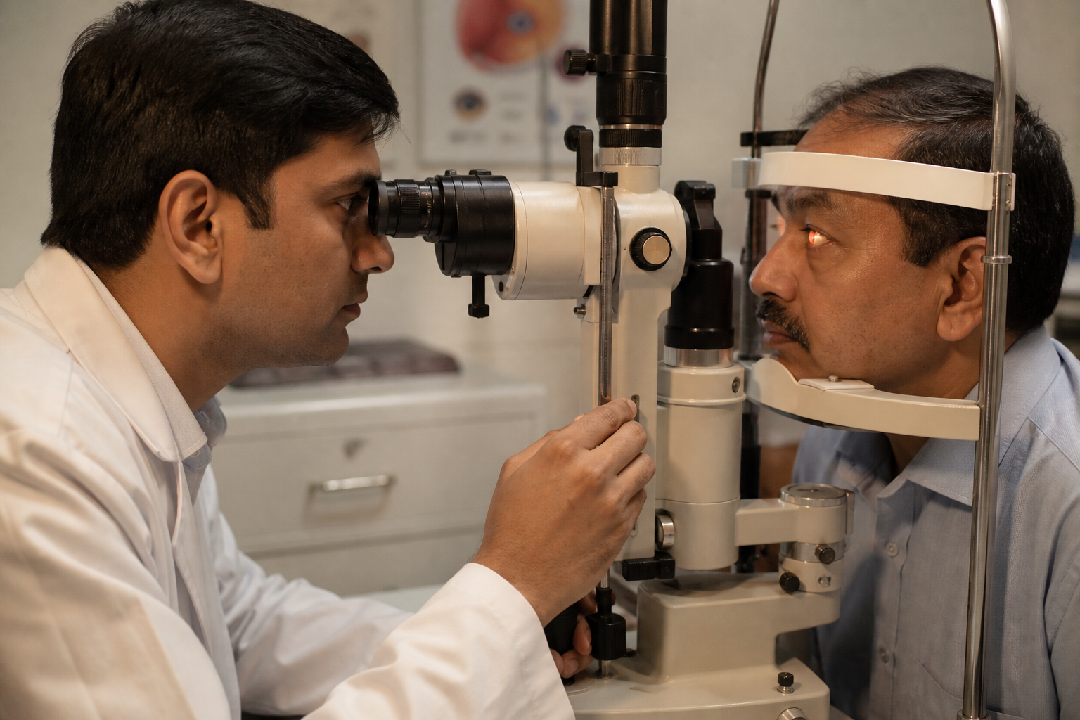
Glaucoma is a group of diseases that slowly damage the optic nerve, which carries visual information from the eye to the brain. Raised intraocular pressure is the main treatable risk, but the nerve can also suffer when blood flow is reduced. Untreated glaucoma can lead to irreversible blindness. Early disease often has no pain, which is why screening matters.
Why it develops
More than one factor may act together. Age and family history raise risk even when the eye feels normal.
Aqueous humour must leave the eye through the angle. When outflow is reduced, pressure rises and the nerve is compressed over time.
Corticosteroids and some dilating drops can raise pressure in susceptible people. Always tell the doctor about steroid use.
The optic nerve needs a steady blood supply. Vascular disease and low perfusion pressure can damage nerve fibres even at modest eye pressure.
How it feels
Open-angle glaucoma is often silent. Angle-closure attacks are sudden and need urgent care.
01
A drop in vision, even if it comes and goes, can signal an acute pressure spike or nerve ischaemia.
02
Halos around lights, especially with ache and nausea, may mean corneal oedema from high pressure.
03
Diplopia is less typical of chronic glaucoma but is recorded when present and investigated.
04
Severe brow or head pain with a red eye and vomiting is an emergency until angle closure is excluded.
05
Unequal pupils or a poorly reactive pupil can accompany acute glaucoma or nerve disease.
Treatment aims to lower intraocular pressure and protect remaining nerve fibres. Care may include prescription drops, oral medicines, laser, surgery, or a combination, according to severity and the type of glaucoma. Vision already lost cannot be restored; the goal is to keep what remains.
01
One of the most accurate methods used to measure intraocular pressure at the slit lamp.
02
A simple, painless measurement of corneal thickness, which is needed to interpret pressure readings correctly.
03
The Humphrey field analyser maps the extent of the visual field so silent side-vision loss can be documented and followed.
04
Optical coherence tomography images and measures the ganglion cell complex and retinal nerve fibre layer without touching the eye.
05
UBM images the anterior segment when the angle, iris and ciliary body need to be seen in more detail.
06
A filtering operation that lowers pressure inside the eye. It remains a gold-standard surgical option in many patients.
07
A valved device opens to drain excess fluid from the eye to an external reservoir when further pressure lowering is required.
08
A tube shunt provides an alternative drainage path when prior surgery has failed or the eye is at high risk.
09
Surgery in children most often uses external trabeculotomy or goniotomy, chosen according to the anatomy of the angle.
On-site equipment
Handheld measurement of intraocular pressure that is quick and comfortable, including for children and post-operative eyes.
High-resolution scans of the optic nerve, RNFL and ganglion cell complex to detect and monitor glaucoma.
Photographs the optic disc so cupping and disc haemorrhage can be compared visit to visit.
Examines the angle pathway and documents the front of the eye.
Used when a laser opening in the iris is anatomically indicated as part of glaucoma care.
Surgical microscope for glaucoma operations performed in this hospital.
Glaucoma FAQs
The main types are:
Risk rises with age. Other factors include raised eye pressure, family history, diabetes, high or low blood pressure, thyroid disorder, eye injury, high refractive error and steroid use.
Avoid activities that strain the eye for several weeks. Use a laxative if you are constipated, so you do not strain while passing stools.
Glaucoma surgery has about a 70 to 90 percent success rate for at least one year in older patients. Individual results depend on the type of glaucoma and previous surgery.
If you are above 40, you should be examined periodically, typically once a year, even if the eyes feel comfortable.
Vision already lost from glaucoma is irreversible. Proper treatment can maintain remaining vision and slow further loss.
to provide comprehensive eye care services to all the sections of society at affordable cost.
Doctors
DR KUMAR SAKET
Cataract, medical retina and trauma specialist
DR MALAY KUMAR DWIVEDI
Pediatric Ophthalmologist, Cataract and Squint specialist
Insurance and schemes
Bring your card and papers. The front desk will explain what is needed before admission.

Ayushman Bharat · PM-JAY
M G Eye Foundation is an empanelled facility under the PM-JAY scheme and welcomes patients seeking eye care under this programme. Kindly consult the front desk for further information prior to admission.
Jharkhand · SEHIS
M G Eye Foundation is associated with the Jharkhand Rajyakarmi Swasthya Bima Yojana (State Employees Health Insurance Scheme / SEHIS). State government employees, pensioners and eligible family members may seek eye care here under this programme. Kindly consult the front desk with your scheme card and papers prior to admission.