
OCT and fundus imaging
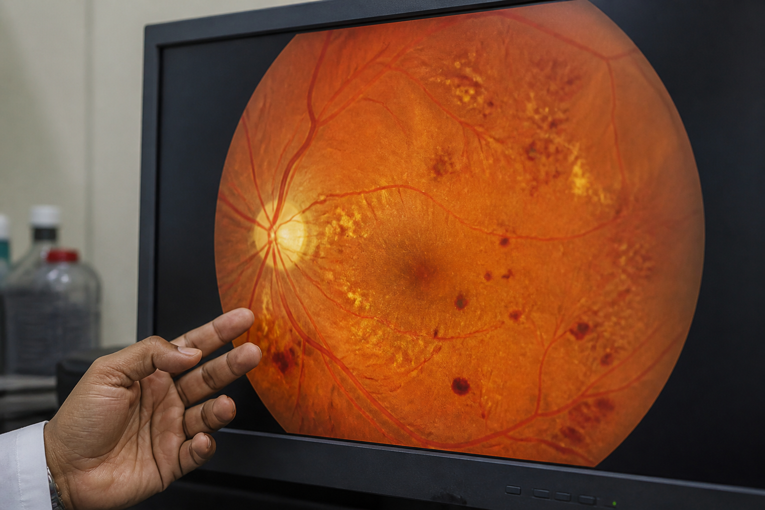
Cross-sectional OCT and colour photographs document diabetic retinopathy, ARMD and macular oedema so treatment and follow-up have a baseline.
Clear Vision. Expert Care. Healthier Eyes.
M G Eye Foundation/Services/Vitreo-retinal Services
The retina lines the back of the eye and converts light into neural signal. Disease of the retinal vessels, macula or vitreous produces blurred, distorted or missing vision. Diabetes, ageing of the macula, retinal vein occlusion, high myopia and injury are common drivers. Untreated, several of these conditions threaten permanent loss.
DR KUMAR SAKET looks after the retina. Injection and laser are done here.
The retina cannot be judged by symptoms alone. Colour fundus photography (Forus) and ZEISS CIRRUS OCT show leakage, oedema, membranes and nerve-fibre change. Medical retina care here includes retinal laser for macular oedema and selected breaks, and intravitreal anti-VEGF or steroid injections for diabetic macular oedema, wet ARMD and vein occlusion. DR MALAY KUMAR DWIVEDI provides ROP screening for premature infants. Fluorescein angiography, ICG angiography, vitrectomy, macular-hole surgery and complex retinal-detachment surgery are tertiary vitreoretinal procedures; if the scan shows that pathway, it is named honestly after the visit.
Medical retina — imaging, laser, injection — and ROP care are delivered here under DR KUMAR SAKET and DR MALAY KUMAR DWIVEDI. Complex vitreoretinal surgery is discussed only when the examination shows it is needed.
Cross-sectional OCT and colour photographs document diabetic retinopathy, ARMD and macular oedema so treatment and follow-up have a baseline.
Laser is used for macular oedema and to seal small retinal breaks, reducing the risk of further leakage or detachment in selected cases.
Medicine is placed in the vitreous cavity for diabetic retinopathy, retinal vein occlusion and wet ARMD, targeting the drivers of fluid and abnormal vessels.
Non-proliferative disease shows microaneurysms, haemorrhages and oedema. Proliferative disease grows fragile new vessels that bleed. Laser, injection and, when needed, surgical referral are sequenced to the stage.
Premature and low-birth-weight babies are screened for abnormal retinal vessel growth. Laser, injection or surgery is planned according to stage.
A torn or detached retina does not heal on its own. Prompt examination is required.
It refers to disease of the vitreous gel and the retina. Medical treatment (laser, injection, imaging) and surgical treatment (vitrectomy) are both used worldwide; we provide the medical-retina and ROP work listed above in this hospital.
According to stage: observation, retinal laser, intra-ocular injections, and surgery in advanced disease. Control of blood sugar, blood pressure and lipids remains part of care.
Monday–Saturday: 10:00 a.m.–6:00 p.m. Sunday: Closed · Call +91 84090 01008
Insurance and schemes
Bring your card and papers. The front desk will explain what is needed before admission.
Ayushman Bharat · PM-JAY
M G Eye Foundation is an empanelled facility under the PM-JAY scheme and welcomes patients seeking eye care under this programme. Kindly consult the front desk for further information prior to admission.
Jharkhand · SEHIS
M G Eye Foundation is associated with the Jharkhand Rajyakarmi Swasthya Bima Yojana (State Employees Health Insurance Scheme / SEHIS). State government employees, pensioners and eligible family members may seek eye care here under this programme. Kindly consult the front desk with your scheme card and papers prior to admission.